Category: Radiology Updates

Radiology Reporting Solutions

Revolutionizing Healthcare: The Power of Radiology Reporting Solutions

In the ever-evolving landscape of healthcare, advancements in technology have significantly transformed the way medical professionals diagnose and treat patients. One such groundbreaking development is the implementation of Radiology Reporting Solutions. These solutions are revolutionizing radiology practices, streamlining workflows, and improving patient care by enhancing the accuracy and efficiency of radiology reports. In this blog, we’ll explore the remarkable impact of Radiology Reporting Solutions in the medical field.

What Are Radiology Reporting Solutions?

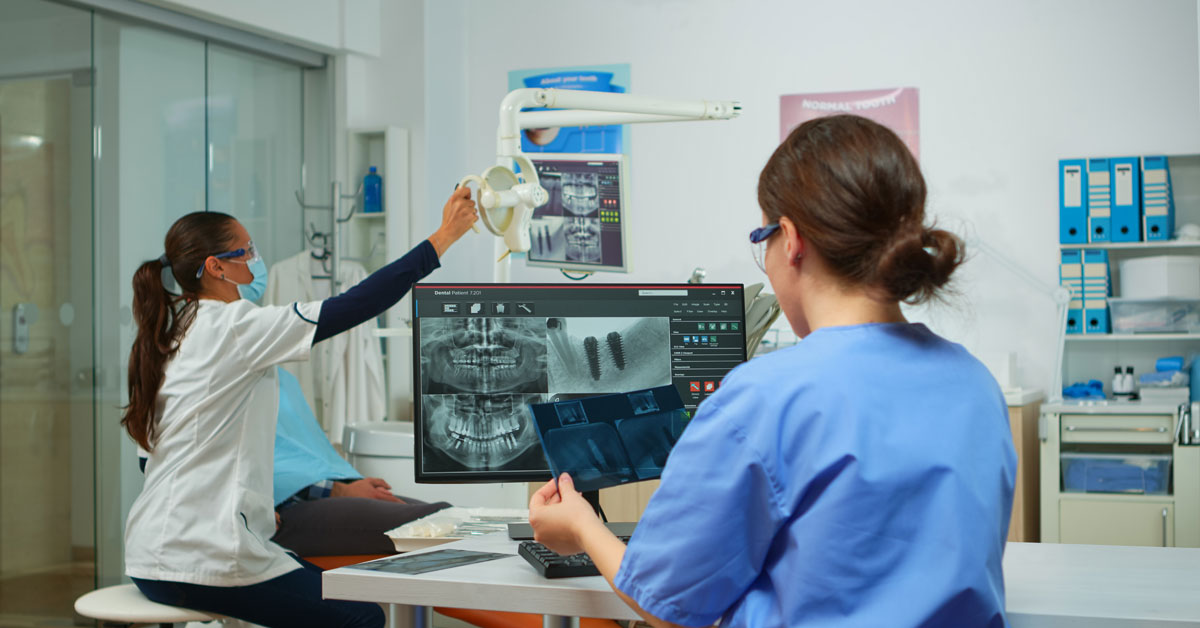
Radiology Reporting Solutions are software systems designed to assist radiologists in generating accurate and comprehensive reports based on medical imaging studies, such as X-rays, MRIs, CT scans, and ultrasounds. These solutions offer a wide range of tools and features that facilitate the interpretation of medical images, making the radiologist’s job more efficient and precise.

Key Benefits of Radiology Reporting Solutions

Enhanced Accuracy: One of the most significant advantages of these solutions is their ability to reduce human error. Advanced algorithms and AI-driven tools assist radiologists in detecting anomalies and abnormalities that might be missed during manual interpretation. This ensures that patients receive more accurate diagnoses and timely treatment.

Efficiency and Workflow Optimization: Radiology Reporting Solutions streamline the reporting process, enabling radiologists to generate reports more quickly and with greater consistency. This efficiency translates to shorter waiting times for patients, reduced administrative overhead, and improved overall workflow in radiology departments.

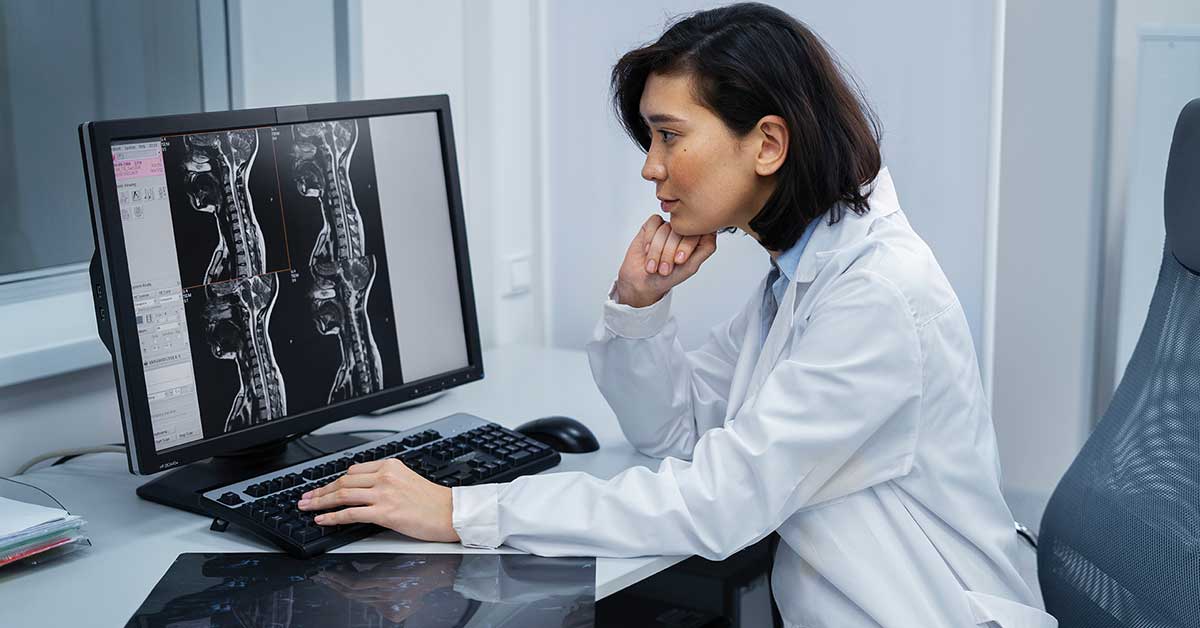
Remote Reporting: With the advent of telemedicine, Radiology Reporting Solutions allow radiologists to work from remote locations, providing expert interpretation and analysis to healthcare facilities and patients regardless of geographical boundaries. This flexibility not only improves patient access to radiology services but also addresses the shortage of radiologists in certain regions.

Structured Reporting: These solutions support structured reporting, which helps standardize the format and content of radiology reports. This consistency enables better communication between healthcare providers, leading to improved patient care and outcomes.

Integration with Electronic Health Records (EHR): Radiology Reporting Solutions seamlessly integrate with EHR systems, allowing for easy access to patient history, previous reports, and other relevant medical information. This integration ensures that radiologists have a comprehensive view of a patient’s health, leading to more informed diagnoses.

Quality Control: Radiology Reporting Solutions often include quality control tools that help maintain high standards in reporting. Radiologists can review and validate reports efficiently, ensuring that the final document is of the highest quality.

The Future of Radiology Reporting

As technology continues to advance, Radiology Reporting Solutions will evolve as well. Expect to see further integration of artificial intelligence and machine learning algorithms, leading to even more accurate and efficient reports. Additionally, these solutions will likely become more patient-centric, allowing patients to access their own reports and images, empowering them to take an active role in their healthcare.

In conclusion, Radiology Reporting Solutions are a game-changer in the healthcare industry. They improve the accuracy, efficiency, and accessibility of radiology services, ultimately leading to better patient care and outcomes. As technology continues to advance, the future holds even more promise for radiologists and patients alike, thanks to these remarkable solutions.

Smoldering Spots in the Brain May Signal Severe MS

Aided by a high-powered brain scanner and a 3-D printer, National Institutes of Health (NIH) researchers found that dark rimmed spots called chronic active lesions may be a sign of more aggressive MS.

The team previously shared instructions for programming lower-powered MRI scanners, found at most clinics, to detect rimmed chronic active lesions. They hope researchers around the world will use the instructions to develop and monitor better diagnostic and therapeutic strategies for MS patients.

Read more: https://www.itnonline.com/content/smoldering-spots-brain-may-signal-severe-ms

Improving efficiency is key to radiology’s future

 Radiology needs to become more efficient and adapt to the shift towards value-based care.

Capitalizing on IT innovation, specifically artificial intelligence (AI), will be important in the coming years as radiology becomes more complex. Physicians will need to set aside fears around AI. AI programs will simply be providing clinical decision support.

Another major trend in healthcare is to move patients away from expensive hospital care into the outpatient environment. Ultimately, the most important step for leaders moving forward in today’s current landscape is to embrace change

Read more: https://www.auntminnie.com/index.aspx?sec=sup&sub=imc&pag=dis&ItemID=134834

Radiologists’ trust of AI is critical for hospital adoption

Today, most radiology departments with AI-enhanced imaging applications have less-than-ideal workflow integration. Standalone AI solutions that require physicians to swivel to a different screen  or log in to separate systems force the physician to be the human data interoperability layer.

Enterprise AI platforms that deliver intersystem interoperability into physicians’ current workflows are an excellent initial step.

With their reputation as early adopters of new technology, radiologists are perhaps best-suited to guide a hospital in developing a long-term, enterprise-wide AI strategy. In addition, with a significant percentage of patient encounters involving imaging studies, radiologists are gaining real-world experience integrating AI into their workflows and demonstrating the utility of AI to clinicians in other specialties.

Source: https://www.auntminnie.com/index.aspx?sec=sup&sub=aic&pag=dis&ItemID=134824

New imaging method reveals the inner workings of a family of light-sensing molecules

Using an innovative new imaging technique, researchers at Weill Cornell Medicine have revealed the inner workings of a family of light-sensing molecules in unprecedented detail and speed. Optogenetics researchers insert genes for light-sensing molecules in neurons or other cells, enabling them to change the cells’ behavior with light pulses.

This work has revolutionized neuroscience and holds potential for treating neurological diseases as well. The more researchers know about light-sensing proteins, the further they’ll be able to push optogenetics.

Read more: https://www.news-medical.net/news/20220127/New-imaging-method-reveals-the-inner-workings-of-a-family-of-light-sensing-molecules.aspx

3 obstacles radiologists must overcome for virtual patient consultations

According to the American Journal of Roentgenology, experts listed the barriers that lie in the way of streamlining virtual radiology consultations. Three key roadblocks are highlighted below.

  • Lack of access to telehealth.
  • Participation of Radiologists and Physicians.
  • Proper reimbursement for virtual visits.

Read more: https://www.healthimaging.com/topics/enterprise-imaging/virtual-radiology-consultations


Researchers are developing a modified MRI protocol to help treat brain hemorrhages

Researchers with the University of Texas Health Science Center at Houston are investigating how different MRI sequences can assist physicians in treating patients with brain hemorrhages. 

With this MRI sequence, the team hopes to see within the hematoma what percentage of the blood is already clotted and what percentage is in the liquid form. It can be determined if patients with mostly clotted blood are less likely to see their blood expand.

Read more: https://www.healthimaging.com/topics/diagnostic-screening/modified-mri-protocol-treat-brain-hemorrhages

Groundbreaking AI Technology Accurately Diagnoses COVID-19 Using Chest X-Rays in Minutes

A pioneering artificial intelligence (AI) technology is capable of accurately diagnosing COVID-19 in just a few minutes.

The groundbreaking program developed by researchers at University of the West of Scotland (UWS; Scotland, UK) is able to detect the virus far more quickly than a PCR test, which typically takes around two hours. It is hoped that the technology can eventually be used to help relieve strain on hard-pressed accident and emergency departments, particularly in countries where PCR tests are not readily available.

Read more:  https://www.hospimedica.com/covid-19/articles/294791287/groundbreaking-ai-technology-accurately-diagnoses-covid-19-using-chest-x-rays-in-minutes.html

Eliminating Lenses: Ghost Imaging Speeds Up X-Ray Fluorescence Chemical Mapping

Researchers have developed a new, focus-free technique for creating chemical maps using x-ray fluorescence. The approach offers fast, high-resolution measurements, which could be useful for analyzing chemical composition for a range of applications in biomedicine.

The new x-ray computational ghost fluorescence approach produces two sets of data for each photon energy — one with the spatial distributions of the input beam and one with the emitted fluorescence measurements. A computer program then puts these data together and overlays all the imaging data from the various photon energies to create a chemical element map of the object.

Read more:  https://scitechdaily.com/eliminating-lenses-ghost-imaging-speeds-up-x-ray-fluorescence-chemical-mapping/

Novel Nuclear Imaging Probe Rapidly Assesses Treatment Response in Gastric Cancer

A novel nuclear imaging probe can measure a gastric cancer patient’s response to treatment within days, marking a potential breakthrough that could enable patients who aren’t responding to treatment to get moved quickly to potentially more effective therapies

Current imaging modalities show tumor size, but it may take weeks before any perceptible change is demonstrated that can indicate whether the treatment is working. There is a need for new imaging approaches so that patients can be scanned within a few days after receiving treatment to see if it’s working or not.

Read more: https://www.hospimedica.com/critical-care/articles/294791225/novel-nuclear-imaging-probe-rapidly-assesses-treatment-response-in-gastric-cancer.html